|
|
Avascular Necrosis of the Talus
General Considerations
- The vascular supply to the talus consists of relatively small vessels combined with poor collateral circulation which predispose the talus to avascular necrosis when there is a disturbance of its blood supply
- The talus is also covered by a large percentage of articular cartilage which is not penetrated by blood vessels
- Cause may be atraumatic or traumatic
- Atraumatic causes include
- Steroids
- Sickle cell disease
- Lupus
- Alcoholism
- Traumatic cause is frequently due to a fracture/dislocation through the neck of the talus
- Risk of AVN of the talus following injury uses the Hawkins Classification
Hawkins Classification for Risk of Avascular Necrosis of Talus |
Type |
Description |
Risk of AVN |
I |
Nondisplaced talar neck fracture |
0%–15% |
II |
Displaced fractures with dislocation or subluxation of subtalar joint |
20%–50% |
III |
Displaced fractures with dislocation or subluxation of both ankle joint and subtalar joint |
~100% |
IV |
Displaced fractures with dislocation or subluxation of subtalar, tibiotalar, and talonavicular joints |
100% |
- The Hawkins sign is visualized as a thin subchondral radiolucent line along all or part of the talar dome best seen on the AP view
- Becomes evident 6–8 weeks after injury
- Its presence indicates an adequate blood supply that can lead to bone resorption and is an indicator used to determine that AVN of the talus will probably not occur
Clinical Findings
- Possibly, increasing pain, limitation of motion and signs of inflammation
Imaging Findings
- Conventional radiography is used most often in following the timeline of talar AVN
- It usually takes between 3-6 months for imaging findings to manifest
- First appears as an area of sclerosis in dome of the talus
- May extend into the body of the talus
- Collapse of the articular surface
- Fragmentation of the talar dome and body
- Due to the processes of reossification and resorption, a thin radiolucent or sclerotic line may outline the edge of the area of avascular necrosis
- Coronal CT is especially helpful in showing the articular surface of the talar dome
- MRI is most sensitive
Treatment
- Goal of fracture treatment in talar neck fractures is to restore neck to its anatomic position
- Closed reduction attempt after first radiographs
- Emergent ORIF for all open/irreducible fxs
- Attempt reconstruction
Prognosis
- Hawkin’s Classification above


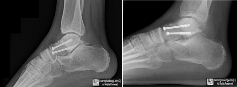
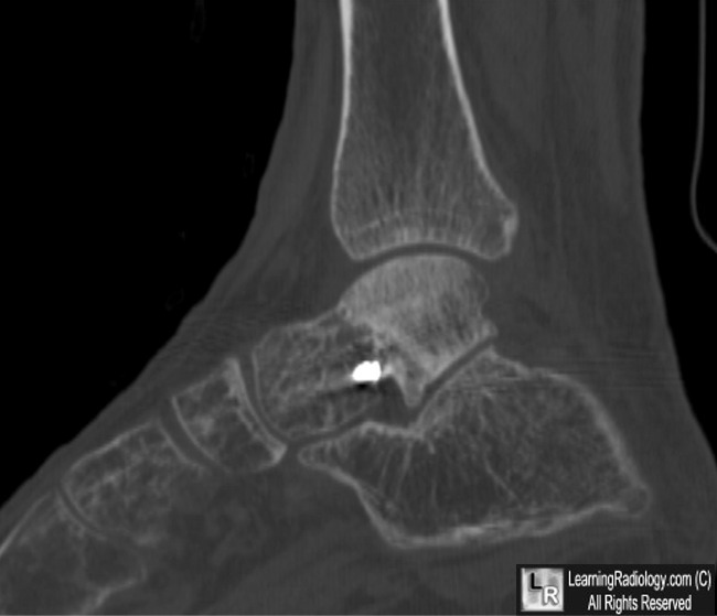
Avascular Necrosis (AVN) of the talus. Upper photos: Lateral radiographs show marked sclerosis of the talar dome and posterior body of the talus (white arrows) relative to the anterior body of the talus (yellow arrows). The patient had a fracture through the talar neck repaired with two screws 6 months earlier. Bottom: Sagittal CT scan through the same area demonstrates the sclerosis of the necrotic bone (white arrow) and the normal talus (yellow arrow).
For these same photos without the arrows, click here and here
For more information, click on the link if you see this icon 
Avascular Necrosis of the Talus: A Pictorial Essay. DH Pearce, CN Mongiardi, VL Fornasier, and TR Daniels. March 2005 RadioGraphics, 25, 399-410.
|
|
|
